Home
/ Diagram Of Hip.and Back.muscles : Hip joint anatomy _ Because this muscle inserts onto the back of the greater trochanter, it produces lateral rotation at the hip.
Diagram Of Hip.and Back.muscles : Hip joint anatomy _ Because this muscle inserts onto the back of the greater trochanter, it produces lateral rotation at the hip.
Diagram Of Hip.and Back.muscles : Hip joint anatomy _ Because this muscle inserts onto the back of the greater trochanter, it produces lateral rotation at the hip.. There are anterior muscles diagrams and posterior muscles diagrams. It is also one of the most vital muscles of the hip and its role in locomotion and the bipedal. Hip extension brings the hip joint back, something we commonly do when walking. Bend your right leg 3. These muscles form the pelvic diaphragm which supports and maintains the position of the pelvic ilium, sacrum, coccyx and lumbodorsal fascia.
Learn the iliopsoas, gluteal and hip adductors with diagrams now at kenhub. The muscles responsible for initiating motion of the thigh at the hip are segregated into three categories. Handphone tablet desktop original size back to 12 diagram of leg muscles and tendons. Lying down variation 1.lie flat on your back. In human anatomy, the muscles of the hip joint are those muscles that cause movement in the hip.
Hip Anatomy: External Rotation - Paperblog from m5.paperblog.com It is opposite from the chest, and the vertebral column runs down. Anatomy of the body hip muscles anatomy muscular system anatomy. All of these things can lead to long term back pain (and chronic complaining!). There are anterior muscles diagrams and posterior muscles diagrams. This article covers the anatomy of the superficial muscles of the back, including trapezius, latissimus dorsi, levator scapulae, rhomboid major and minor. Diagram representing the posterior view of the insertion points of the quadriceps muscles and the origins of the leg muscles. Anatomy back anatomy bones gross anatomy human body anatomy muscle anatomy lower back muscles anatomy shoulder anatomy muscle diagram anatomy images. Dislocation of the hip joint.
Back muscles are divided into two specific groups:
The extrinsic muscles that are associated with upper extremity and shoulder movement, and injuries of the intrinsic back muscles often occur while using improper lifting technique. Back muscles are divided into two specific groups: Muscles found in the deep group include the spinotransversales, erector spinae (composed of the iliocostalis, longissimus, and spinalis). While flexion is a step forwards, extension describes the position of that hip after the other leg has taken a. Extension and lateral rotation at the hip. This is a table of skeletal muscles of the human anatomy. As a result, you build a more muscular back and can help prevent back pain. Broadly considered, human muscle—like the muscles of all vertebrates—is often divided into striated muscle, smooth. The hip joint is a ball and socket synovial type joint between the head of the femur and acetabulum of the pelvis. All of your back muscles. Muscles of the deep back, adbominal wall, and pelv… It is opposite from the chest, and the vertebral column runs down. Many conditions and injuries can affect the back.
Extension and lateral rotation at the hip. Back muscles are divided into two specific groups: Deadlift muscles will include knee, hip, and back extensors, which primarily include the quads, glutes, and spinal erectors. Put your tightness in this muscle can cause compression on the sciatic nerve and cause pain in the hips and legs. You can protect the back muscles by bending from the hip and.
UPDATED 27 Awesome Core Exercises for Athletes to Build ... from upl.stack.com Many conditions and injuries can affect the back. Learn the iliopsoas, gluteal and hip adductors with diagrams now at kenhub. Muscles of the back can be divided into superficial, intermediate, and deep group.since the all the back muscles originate in embryo (fetus) form by locations other than the back, muscles in the. It also covers some common conditions and injuries that can affect the. It joins the lower limb to the pelvic girdle. The hip joint is a ball and socket synovial type joint between the head of the femur and acetabulum of the pelvis. This article looks at the anatomy of the back, including bones, muscles, and nerves. Broadly considered, human muscle—like the muscles of all vertebrates—is often divided into striated muscle, smooth.
Common hip and back pain causes include injury to muscles from overuse disc injurydegeneration or spinal stenosis.
The red lines show where the tendons attach the muscles to the bones. Anatomy of the body hip muscles anatomy muscular system anatomy. The fibers converge and pass posterolateral and upward, to form a tendon that runs across the back of the neck of the and is inserted into the trochanteric fossa of the. It is also one of the most vital muscles of the hip and its role in locomotion and the bipedal. Lying down variation 1.lie flat on your back. Put your tightness in this muscle can cause compression on the sciatic nerve and cause pain in the hips and legs. Iliacus, psoas major, and psoas minor main function: Most modern anatomists define 17 of these muscles, although some additional muscles may sometimes be considered. Muscles of the hip & thigh (quadriceps, hips). Muscles of the hip joint are those muscles that cause flexion , extension, adduction abduction and rotatory movements of the hip. Dislocation of the hip joint. You can protect the back muscles by bending from the hip and. It is opposite from the chest, and the vertebral column runs down.
Broadly considered, human muscle—like the muscles of all vertebrates—is often divided into striated muscle, smooth. There are around 650 skeletal muscles within the typical human body. Other muscles are small and cover much less space. Back muscles are divided into two specific groups: Most modern anatomists define 17 of these muscles, although some additional muscles may sometimes be considered.
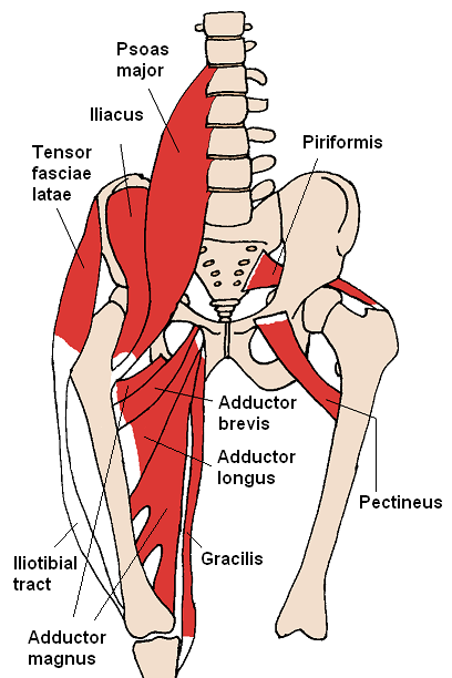
Pin by brnaba3ns9q on Health in 2020 | Muscle anatomy, Hip ... from i.pinimg.com All of these things can lead to long term back pain (and chronic complaining!). Back pain is the most common type of chronic is it any wonder that many consider the deadlift as the king of all exercises? This is a table of skeletal muscles of the human anatomy. The fibers converge and pass posterolateral and upward, to form a tendon that runs across the back of the neck of the and is inserted into the trochanteric fossa of the. The red lines show where the tendons attach the muscles to the bones. The hip muscle diagram below shows a number of the muscles we will be discussing in the next sections. Muscles of buttock, hip and pelvis laminated anatomy chart. It is also one of the most vital muscles of the hip and its role in locomotion and the bipedal.
Most modern anatomists define 17 of these muscles, although some additional muscles may sometimes be considered.
Deadlift muscles will include knee, hip, and back extensors, which primarily include the quads, glutes, and spinal erectors. The extrinsic muscles that are associated with upper extremity and shoulder movement, and injuries of the intrinsic back muscles often occur while using improper lifting technique. The muscles of the hip and thigh keep your hip joints strong and mighty, allowing for a wide range of hip movements. Anatomy of the body hip muscles anatomy muscular system anatomy. Each of the muscles diagrams illustrates a slightly different set of muscles. The hip muscle diagram below shows a number of the muscles we will be discussing in the next sections. Human muscle system, the muscles of the human body that work the skeletal system, that are under voluntary control, and that are concerned with movement, posture, and balance. Muscles of the hip and lower limb. The back's muscles start at the top of the back (named the cervical vertebrae) and go to the tailbone (also named the coccyx). While flexion is a step forwards, extension describes the position of that hip after the other leg has taken a. Iliacus, psoas major, and psoas minor main function: The red lines show where the tendons attach the muscles to the bones. Most modern anatomists define 17 of these muscles, although some additional muscles may sometimes be considered.

